Question 1: Describe the lesion
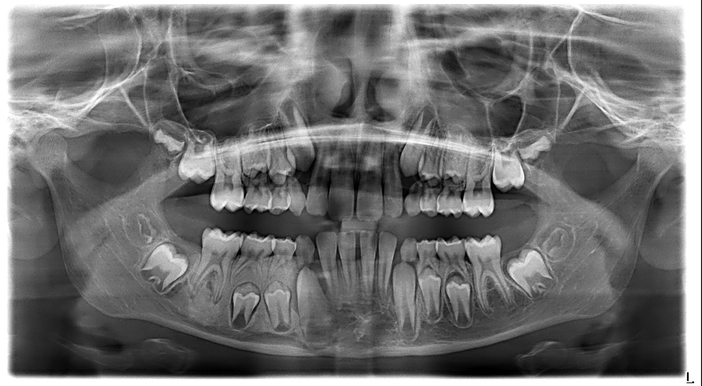
On the right side of the mandible, a radiopaque, sclerotic, and homogeneous lesion is observed, extending from the distal aspect of the unerupted canine to the mesial aspect of the germ of the second molar and from the alveolar ridge down to the inferior alveolar nerve canal.
The lesion has somewhat ill-defined borders, but no expansion, thinning, or perforation of the cortical plates is observed.
The premolar tooth germs and the mandibular canal are displaced toward the lower border of the mandible.
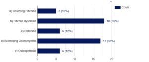
Question 2 Which of the following would you include in your differential diagnosis? (Select one or more correct answers).

The correct answers are: Fibrous Dysplasia and Sclerosing Osteomyelitis.
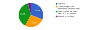
Question 3: What, in your opinion, are the next steps the patient should take in order to reach to a final diagnosis and receive appropriate treatment for this condition? (Select only one).


