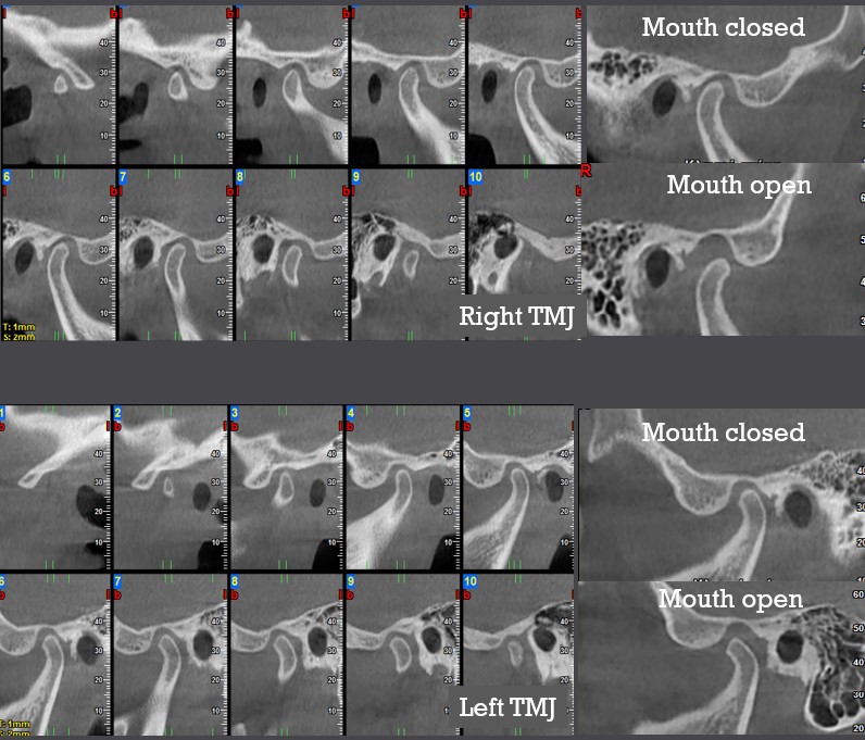
TMJ Internal derangement .
- Internal derangement of the temporomandibular joint (TMJ) is a condition in which the articular disc has been displaced from its original position on the condylar head. The two most commonly observed conditions are: Disk displacement with reduction and Disk displacement without reduction.
- Disc displacement with reduction
- In disc displacement with reduction, the articular disc with the mouth closed has been displaced anterior to the condylar head. It may also be displaced medially or laterally. The disc remains in this position as long as the mouth is closed. When the mouth is opened, the disc is re-situated on the condylar head. The movement of the disc onto and off the condylar head may result in a clicking sound.
- Disc displacement without reduction
- With the mouth closed the articular disc has been displaced anterior to the condylar head and when the mouth is opened it does not reduce into it’s normal position, resulting in limiting opening (locking).
- The two most effective imaging methods for diagnosing TMJ internal derangement, are: MRI and CBCT.
- Imaging Characteristics
- The condyle, the glenoid fossa and the anterior articular eminence do not show osseous lesions or changes.
- In disk displacement with reduction with the mouth closed, in CBCT the condyle is placed in a posterior position into the glenoid fossa. In MRI the disc is visualized displaced anteriorly to the condylar head.
- In disk displacement without reduction with the mouth closed, in CBCT the condyle is placed in a posterior position into the glenoid fossa. In MRI the disc is totally displaced anteriorly to the condylar head.
- With the mouth open in disk displacement with reduction, in CBCT the condyle is brought below or anteriorly the articular eminence. In MRI the disc is depicted in a normal position on the condyle.
- With the mouth open in disk displacement without reduction, in CBCT the condyle is brought posteriorly to the articular eminence or remains into the glenoid fossa. In MRI the disc is visualized situated between the anterior surface of the condyle and the articular eminence.


