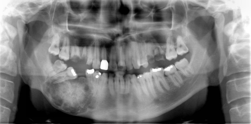
Cemento-Ossifying Fibroma (2nd stage) .
- Benign fibro-osseous neoplasm. It is difficult to differentiate the calcified tissue within the lesion between bone and cementum.
- Most commonly in the third and fourth decade of life.
- It is usually located in the premolar/molar area of the mandible.
- Three stages of development: radiolucent – mixed – radiopaque.
- In early stages it appears as unilocular, well-defined radiolucency.
- As the lesion grows radiopaque foci develop within the radiolucency.
- As the lesion matures it becomes radiopaque. The radiopacity is surrounded by a thin radiolucent ring.
- Generally is asymptomatic unless it grows large enough.
- It can cause displacement of the adjacent teeth. Root resorption is uncommon.


