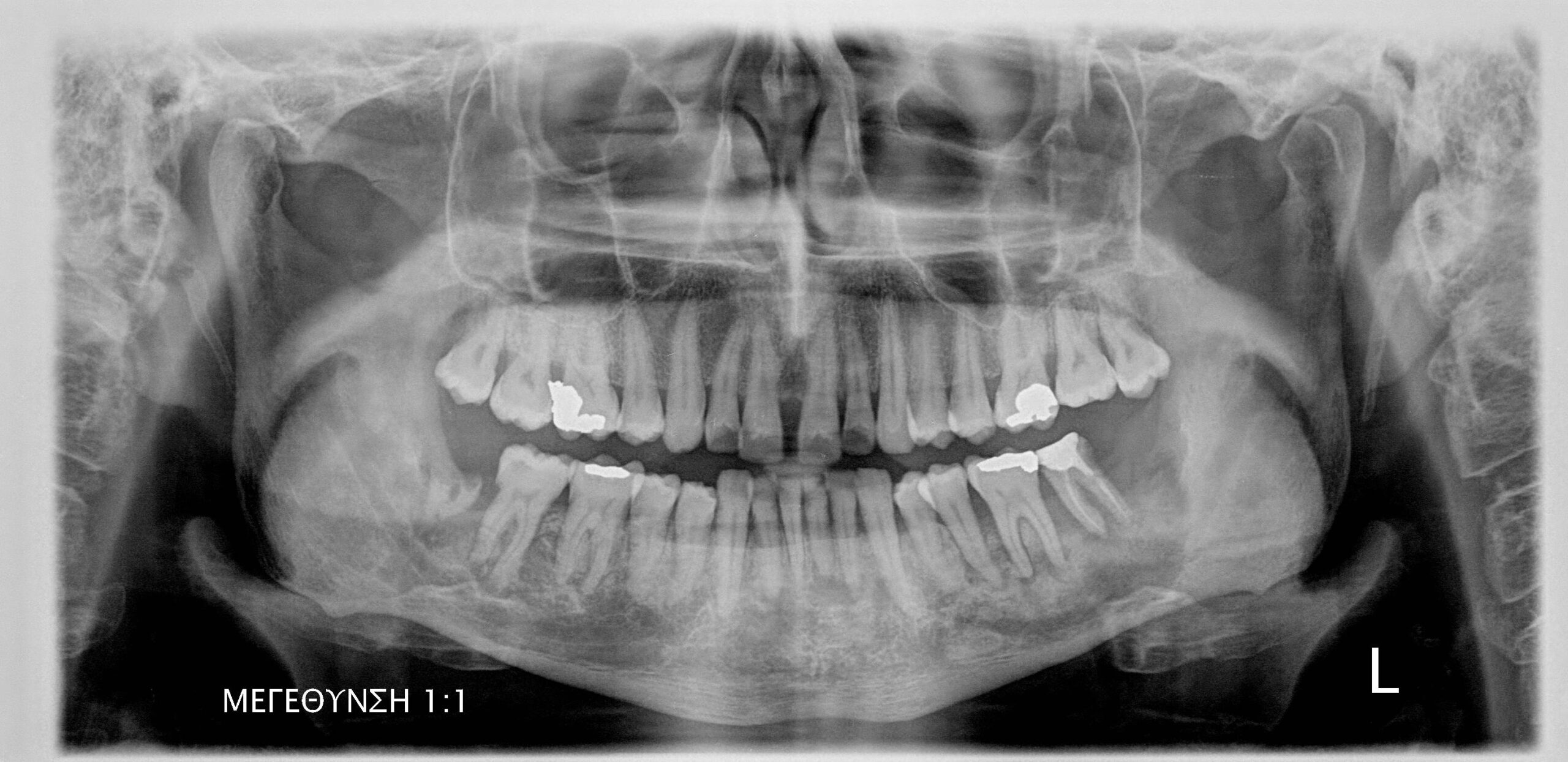
Osteoradionecrosis of the jaws .
- Osteoradionecrosis of the jaw is one of the most serious complications in patients who have undergone radiotherapy for head and neck cancer. It is characterized by the presence of necrotic bone that does not heal after 3 months, typically in patients who have received radiation doses exceeding 60 Gy.
- The mandible is usually affected.
- The diagnosis of osteoradionecrosis is mainly based on the patient’s history of head and neck radiation. Clinically, it usually presents with pain and exposed necrotic bone.
- The preferred imaging modalities for diagnosing and assessing bone lesions are computed tomography (CT), and cone beam computed tomography (CBCT).
- Radiographic features: A diffuse osteolytic lesion with ill-defined and moth-eaten borders is observed, causing destruction and perforation of the cortex. Occasionally bone sequestrion within the osteolytic lesion is observed resulting in a mixed (radiolucent/radiopaque). In advanced stages, the lesion may lead to pathological fractures of the jaw.


