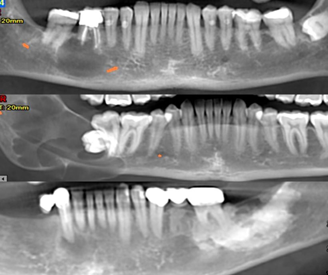
For the successful differential diagnosis, the lesion must first be classified according to its radiographic/imaging characteristics. This classification allows the grouping of pathological conditions with similar radiographic appearance and consequently the limitation of the list of the diseases, for establishing the differential diagnosis.
The classification of bone lesions into large groups with common radiographic characteristics is performed based on the following criteria:
01. Internal structure of the lesion
The relative degree of radiolucency or radiodensity of a lesion compared to the surrounding normal bone varies and depends on the nature of the pathological process.
02. Size and location of the lesion
The size and the location of a lesion in relation to the teeth or to anatomic structures, provide significant information about the type of the underlying condition.
03. Shape of the lesion
The shape can provide information about the extent and the rate of growth of the lesion.
04. Borders of the lesion
The radiographic borders are somewhat indicative of the nature of the lesion and can aid in the differential diagnosis between benign and malignant or inflammatory conditions.
05. Effects on the surrounding bone
Important features when examining a radiograph are the structure of bone trabeculation and the effect of the lesion to the Lamina Dura or to other anatomical landmarks.
06. Effects on the bony cortex
The effect of a lesion to the bony cortex can provide information about the nature of the pathological condition.
07. Effects on the adjacent teeth
The effect of a lesion on the adjacent teeth varies according to the nature of the procedure. A lesion may have no effect on the adjacent teeth, or may lead to displacement or resorption of the roots.
08. Periosteal reactions
A lesion may cause periosteal reactions and this can provide information about the nature of the underlying disease.

AUTHORS/ADMINISTRATORS
Prof. KOSTAS TSIKLAKIS, DDS, MSc, PhD
Professor Emeritus, School of Dentistry,
National and Kapodistrian University of Athens, Greece
Dr. KOSTAS SYRIOPOULOS, DDS, PhD
Oral and Maxillofacial Radiologist
Website Editorial Team
COMMUNICATION SPONSORS
Copyright © 2025 DDMFR. All Rights Reserved. Designed & Developed by Web Idea


