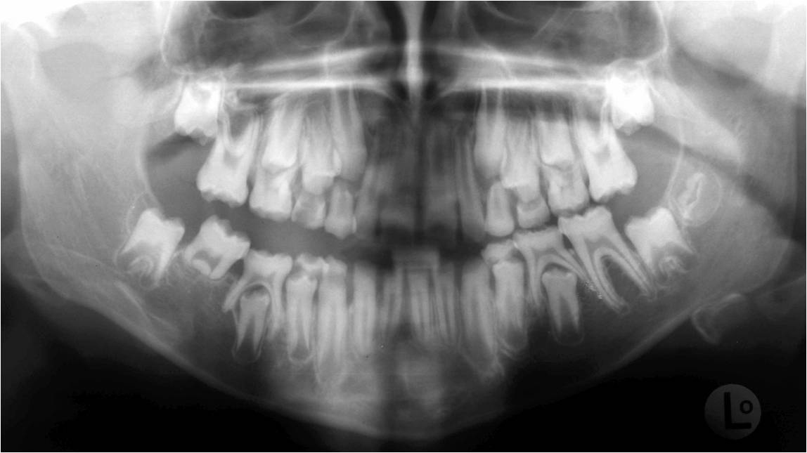
Teeth structure .
- Amelogenesis Imperfecta: is a congenital disorder which presents with a rare abnormal formation of the enamel which is unrelated to any systemic or generalized condition Amelogenesis imperfecta is mainly divided to four types: Hypoplastic (type I); Hypomaturation (type II); Hypocalcified (type III); and Hypomaturation /hypoplasia/ Taurodontism (type IV). Identification of amelogenesis imperfecta is made primarily by clinical examination. The radiographic features include: a square crown, thin radiopaque layer of enamel, low or absent cusps and open contacts between teeth.
- Dentinogenesis Imperfecta: is a genetic disorder involving primarily the dentin, although the enamel may be thinner than normal. Type I is associated with osteogenesis imperfecta with skeletal anomalies. Type II is similar to type I but only affects the dentin without any skeletal defects. The radiographic features include: obliteration of the pulp, short roots, bulbous crown with a marked constriction at cementoenamel junction (tulip teeth).
- Regional Odontodysplasia (ghost teeth): is a relatively rare condition in which both enamel and dentin are hypoplastic or hypocalcified. This condition affects only a few adjacent teeth in one quadrant. The radiographic features include: pulp chambers are large, roots are short, enamel and dentin are very thin. These teeth have been described as having a ‘ghostlike’ appearance.


