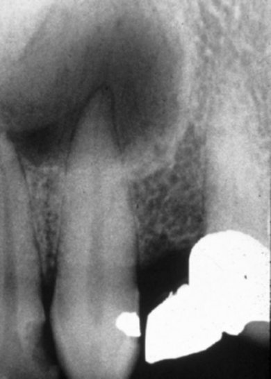
Radicular cyst .
It is an odontogenic inflammatory cyst associated with the apex of a tooth with nonvital pulp.
It originates from epithelial cells of Malassez locating in the periodontal ligament of a nonvital tooth. The epithelial cells under stimulation by the inflammatory products proliferate resulting in cystic degeneration.
It is the most common cyst that develops in the jaws (55% to 70% of al cystic lesions). It’s greater dimension usually does not exceed 1.5-2 cm, but there are cases where it may reach up to 5 cm.
It has a similar radiographic appearance to the periapical granuloma, but is larger in size.
Radiographically appears as a well-defined radiolucency and most often is surrounded by a sclerotic border.
Large radicular cysts may cause expansion, thinning and possibly perforation of the cortical plates of the jaws.
The apical portion of the tooth enters the radiolucency and loss of the lamina dura is observed.
In large radicular cysts displacement and resorption of the roots of the adjacent teeth may be apparent.


