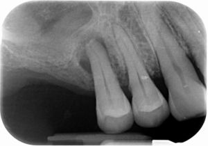
Periapical Abscess .
Periapical abscess clinically is distinguished into acute and chronic. It is an inflammatory process which develops in the periapical region of a tooth with necrotic pulp or in a tooth with incomplete endodontic treatment.
Etiologically, it is divided into primary and secondary. The primary abscess is always acute and initially does not give radiological findings. As primary abscess develops, an increased periodontal ligament space, around the apical part of the root is observed. After 7-10 days a periapical radiolucency with somehow ragged borders is usually apparent in the radiograph.
Secondary periapical abscess develops in a pre-existing apical lesion (granuloma or cyst) and may be acute or chronic. The radiographic image is related to the preexisting entity and discontinuity of the periodontal ligament and lamina Dura is a common radiographic finding.


