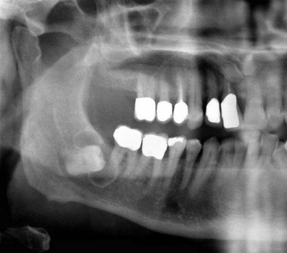
Dentigerous (follicular) cyst .
- It is the second most common type of cyst found in the jaws, following the radicular cyst and is formed around the crown of an unerupted tooth.
- It develops due to the accumulation of fluid between the remnants of the enamel organ (reduced enamel epithelium) and the crown of the tooth.
- Most common sites: mandibular third molars followed by maxillary canines and maxillary third molars.
- Denigerous cysts are found at all ages, but most often in young people from 10 to 30 years old.
- The lesion is usually asymptomatic.
- Radiographically, it appears as a unilocular radiolucency with well-defined and often corticated borders, associated with the crown of an unerupted tooth.
- Large dentigerous cysts surround the entire tooth, which is often displaced and tend to expand the cortical plates of the jaws (usually on the buccal side).
- Eruption cyst is a term to describe a dentigerous cyst in children when it is located in the soft tissues overlying the unerupted tooth.
- Ameloblastomas or squamous cell carcinomas have been reported to arise in the epithelial lining of a dentigerous cyst.


