• Home
  • Lesions of the Jaws
    ▼
    • Basic principles of Differential Diagnosis
    • Radiolucencies (Οsteolytic)
    • Mixed (osteolytic and osteoblastic)
    • Radiopacities (osteoblastic)
  • TMJ
  • Paranasal sinuses
  • Dental Abnormalities
  • Implantology
  • Endontology
  • Members only
  • Contact
  • English
  • Ελληνικά
    • Login
    • Register
    • Home
    • Lesions of the Jaws
      • Basic principles of Differential Diagnosis
      • Radiolucencies (Οsteolytic)
      • Mixed (osteolytic and osteoblastic)
      • Radiopacities (osteoblastic)
    • TMJ
    • Paranasal sinuses
    • Dental Abnormalities
    • Implantology
    • Endontology
    • Members only
    • Contact
    • English
    • Ελληνικά
    • Periapical
      • Periapical Cemento-Osseous Dysplasia 3rd stage
      • Tori and exostoses
      • Condensing osteitis
      • Cemento-Ossifying Fibroma (3rd stage)
      • Hypercementosis
      • Cementoblastoma
      • Foreign body (periapical)
    • Focal
      • Idiopathic osteosclerosis (dense bone island)
      • Osteoma and exostoses
      • Fibrous dysplasia (radiopacity)
      • Odontoma (radiopacity)
      • Soft tissue calcifications
      • Foreign bodies (focal)
      • Root remnants
      • Socket sclerosis
    • Multiple & generalized
      • Multiple impacted teeth
      • Florid cemento-osseous dysplasia (radiopacity)
      • Foreign bodies (multiple)
      • Sclerosing Osteomyelitis
      • Paget’s disease (stage 3)
      • Soft tissue calcifications
      • Osteogenesis imperfecta
      • Osteopetrosis

    Sclerosing Osteomyelitis .

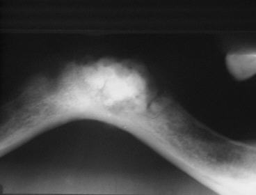
    • It is a rare, chronic, non-pyogenic form of osteomyelitis  that primarily affects the  mandible. It is characterized by inflammation, sclerosis (hardening), and thickening of the bone due to a prolonged infection.
    • Radiographically, it appears as a diffuse radiopacity with ill defined borders.

    Differential Diagnosis

    • Fibrous dysplasia (radiopacity)
    • Idiopathic osteosclerosis (dense bone island)
    • Osteoma and exostoses

    Case 1

    A case of sclerosing osteomyelitis

    Case 2

    Case of sclerosing osteomyelitis

    AUTHORS/ADMINISTRATORS
    Prof. KOSTAS TSIKLAKIS, DDS, MSc, PhD

    Professor Emeritus, School of Dentistry,
    National and Kapodistrian University of Athens, Greece

    Dr. KOSTAS SYRIOPOULOS, DDS, PhD

    Oral and Maxillofacial Radiologist

    Website Editorial Team

    COMMUNICATION SPONSORS

    Vatech
    Dentsply Sirona
    • Πολιτική Απορρήτου
    • Φόρμα Επικοινωνίας
    • Privacy Policy
    • Contact us

    Copyright © 2025 DDMFR. All Rights Reserved. Designed & Developed by Web Idea