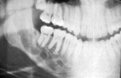
Hemangioma, central .
- It is a rare benign tumor which is found mainly in the spine, the skull and rarely in the jaws.
- It may occur at any age, but is more common in adolescents.
- Radiographically it appears as a multilocular radiolucency (soap bubble or honeycomb appearance). Usually the lesions of the jaws are connected to the large vessels of the neck.
- Root resorption of the adjacent teeth are common. Developing teeth may be displaced or erupt earlier.
- When the lesion involves the inferior dental canal, the canal may be enlarged.
- Aspiration of the lesion and angiography are important diagnostic tools.


